Mesothelioma Radiology
Radiology is the use of X-rays and other technologies to diagnose and treat disease. It plays an important role in mesothelioma diagnosis and treatment. Radiology includes diagnostic imaging such as X-rays, CT scans, MRIs and PET scans, as well as interventional radiology for certain procedures.
How Is Radiology Used to Diagnose Mesothelioma?
Radiology plays a crucial role in diagnosing mesothelioma. Diagnostic scans for mesothelioma can include X-rays, computed tomography, magnetic resonance imaging and positron emission tomography. Each of these techniques has different strengths and weaknesses when it comes to identifying malignant mesothelioma.
Imaging scans are also used for mesothelioma staging. Accurate staging requires determining the tumor’s size and location, whether lymph nodes are involved and if cancer cells have spread to other parts of the body.
I believe we all have scanxiety in the beginning. One thing I came to realize was that it was to be expected. We had never been in that type of situation before and we didn’t know what to think.
Because it can find cancer cells throughout the body, PET/CT scans are a valuable tool for determining the stage of mesothelioma. Research also shows that PET/CT is effective for tracking response to treatment. It can show whether chemotherapy or other treatments are killing cancer cells throughout the body.
Radiology imaging is also used to guide needles for biopsy — the only way to definitively diagnose mesothelioma. Using X-rays or CT, your doctor can precisely guide a needle to obtain tissue samples from suspected tumors. This is especially important for very small tumors, tumors that are difficult to reach or in people who are not candidates for a surgical biopsy.
- X-rays provide a 2D image that can show tumors, fluid buildup and other signs of disease.
- CT scans use a machine that takes many X-rays to produce 3D images of cross-sections of the body.
- MRI uses a strong magnetic field to produce a 3D image of soft tissues in the body. It can detect mesothelioma earlier than X-ray or CT scans.
- PET/CT uses a CT scan taken after injecting a radioactive dye into the bloodstream to find cancer cells in the body.
Can Radiology Detect All Mesothelioma Types?
Imaging scans can identify all 4 types of mesothelioma: Pleural, peritoneal, pericardial and testicular. However, some types of imaging scans are better suited than others for visualizing mesothelioma in different locations in the body.
- Pleural and Pericardial Mesothelioma: Tumors, fluid accumulation and other abnormalities can be seen with chest X-rays.
- Peritoneal and Testicular Mesothelioma: X-ray isn’t very good, however, for identifying abnormalities in soft tissue, such as the gut and the testicles.
CT scans are the primary type of imaging used to detect mesothelioma. They can provide a cross-section of the chest with much more detail than an X-ray and see tumors or other abnormalities in the abdomen and groin. MRI scans are similar to CT but offer higher resolution and can find small tumors, lymph nodes and metastases that are difficult to see with CT.
The most versatile imaging technique for mesothelioma diagnosis is PET/CT imaging. It can detect very small tumors anywhere in the body and can be used to help diagnose every type of mesothelioma. However, PET scans are very expensive and aren’t available everywhere.
What Does Mesothelioma Look Like on an X-Ray?

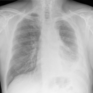
On a chest X-ray, pleural or pericardial mesothelioma tumors appear as wispy white areas around the lungs, while calcified tumors appear bright white. Bones appear white and healthy lungs are dark. Most abnormalities appear as lighter areas that are hazy or solid.
Tumors and scarring may distort chest anatomy. Compressed lungs or a raised diaphragm can be visible on an X-ray.
X-rays are 2D, making it hard to determine if a tumor is in the lung, pleura or the mediastinum around the heart. Additionally, X-rays don’t clearly show peritoneal or testicular mesothelioma. CT, MRI and PET/CT scans offer more detailed images for all mesothelioma types.

Radiology-Guided Procedures for Mesothelioma
Doctors can use radiology imaging to see inside the body while performing procedures for diagnosis and treatment. With ultrasound, CT or MRI, doctors can precisely place needles and similar instruments into the body.
They can reach tumors that are very small or lie deep within the body with very long needles without the need for surgery. These procedures include fine-needle biopsies, tumor ablation and procedures to drain fluid.
- Ablation: Using CT or ultrasound, a needle-like probe is precisely placed into tumors. Small tumors can be destroyed with heat, cold, lasers, microwaves or radio waves.
- Biopsy: Fine-needle biopsies collect tissue samples from tumors. Doctors use ultrasound or CT to precisely place a needle into a tumor to collect a tissue sample.
- Catheter placement: CT scanning can guide the placement of a catheter or port for drug infusions or chemotherapy injected directly into the blood vessels feeding a tumor (transarterial chemoperfusion).
- Drainage: Fluid around the heart or other hard-to-reach areas can be drained using a precisely placed needle. CT or ultrasound is necessary to guide the needle when tumors and scarring distort the normal anatomy.
One of the most exciting advances in interventional radiology treatments for mesothelioma is transarterial chemoperfusion. This procedure uses CT-guided placement of needles to deliver chemotherapy drugs directly into the arteries that are providing blood to a tumor. This allows the chemotherapy drugs to attack the cancer while minimizing unwanted side effects.
Radiology-guided procedures do carry some risk of bleeding, infection and injury. However, these procedures are much less risky than invasive surgery and require less time for recovery. This is very important for people who cannot undergo surgery because of their health.
What Is Interventional Radiology?
Interventional radiology is the use of radiology to perform procedures that not only diagnose but also treat diseases such as mesothelioma. This includes imaging-guided procedures as well as radiation treatments.
New radiation treatments and other interventional radiology procedures are becoming increasingly popular in mesothelioma treatment because of their effectiveness and relative safety. Transarterial chemoperfusion, for example, involves identifying specific blood vessels that feed tumors. It delivers high doses of chemotherapy drugs to stop cancer growth without exposing the rest of the body.
We were pleasantly surprised to find that [transarterial chemoperfusion] doesn’t come with the same side effects of traditional intravenous chemotherapy. To see these promising results with so few side effects means we are able to make a positive impact on quality of life for these patients.
Radiation treatments for mesothelioma can improve survival, reduce pain and prevent metastasis throughout the body. They can include external-beam radiation, intensity-modulated radiation therapy or 3D conformal radiation therapy.
New techniques minimize exposure of healthy tissue to harmful radiation using smaller amounts of radiation aimed at the tumor from different directions. Clinical trials are developing cutting-edge therapies to enhance interventional radiology techniques and improve the lives of mesothelioma patients.
Recommended Reading


