Cytokeratin 5 & 5/6 and Mesothelioma
When found in high quantities in tissue samples, cytokeratin 5 and 5/6 proteins can act as markers that help pathologists detect cancers like mesothelioma. Testing for these markers can help people exposed to asbestos get an earlier and more accurate mesothelioma diagnosis.

What Is Cytokeratin 5 and 5/6?

Cytokeratin 5 and 5/6 are proteins normally found in your body. They help build and support certain cells, including those that line your lungs. Finding unusually high levels of these proteins in tissue samples, however, can be a sign of diseases like cancer.
It’s more common to find these high levels with some kinds of cancer – like mesothelioma – than with other types. This makes cytokeratin 5 and 5/6 helpful indicators or biomarkers for mesothelioma. This can help pathologists tell the difference between mesothelioma and similar cancers.
For example, adenocarcinoma has similar symptoms as pleural mesothelioma. Adenocarcinoma starts in the glands and can spread to the lungs. Despite some similarities between these cancers, elevated cytokeratin 5 and 5/6 isn’t common with adenocarcinoma. Finding these markers can help rule adenocarcinoma out. And it could indicate the patient has mesothelioma.
How Are Cytokeratin 5 and 5/6 Used to Diagnose Mesothelioma?
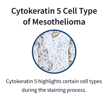
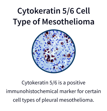
Cytokeratin markers are part of a multi-step process to diagnose mesothelioma. Tissue samples are collected and stained with antibodies, which can reveal different key proteins (or immunohistochemical markers).
A 2022 study in the medical journal Medicina showed cytokeratin was among the most helpful biomarkers for diagnosis. Presence of cytokeratin 5 and 5/6 is a hallmark of mesothelioma. Research indicates it offers up to 90% accuracy in identifying epithelioid mesothelioma cases.
Diagnostic Process Using Cytokeratin
- Tissue biopsy: A small tissue sample is collected, often from the lining of the chest (pleura) or abdomen (peritoneum).
- IHC testing: Pathologists apply antibodies to the tissue sample to detect cytokeratin 5 and 5/6 proteins.
- Marker analysis: Confirming the presence of cytokeratin 5 and 5/6 can help distinguish mesothelioma from other cancers.
- Additional markers: Looking for the presence of other markers as well, such as calretinin or WT1, increases diagnostic accuracy.
Finding cytokeratin 5 and 5/6 isn’t enough on its own to confirm a mesothelioma diagnosis, though. Pathologists also look for other markers as well like calretinin or WT1. Finding all of these markers together can help better distinguish between different types of cancer.
This precise approach helps avoid misdiagnosis. This type of cancer is aggressive, so early and accurate identification is important. A confirmed diagnosis as soon as possible allows people to begin treatment earlier.
The Importance of Cytokeratin in Early Mesothelioma Diagnosis

Using cytokeratin 5 and 5/6 in the diagnostic process helps ensure an accurate diagnosis. A misdiagnosis can lead to initially being given incorrect or unnecessary treatments. An accurate diagnosis can help people receive prompt care tailored to their needs.
Confirming mesothelioma early is vital for high risk patients, such as those with known asbestos exposure. Beginning treatment before the cancer spreads offers a better chance of controlling it.
Early-stage mesothelioma responds better to treatment. People also have more options at earlier stages of mesothelioma, such as joining clinical trials for innovative therapies.

Limitations of Cytokeratin 5 & 5/6 Testing for Mesothelioma
While cytokeratin testing is highly effective, there are challenges. In rare cases, there are overlapping markers. This means mesothelioma isn’t the only cancer where cytokeratin 5 and 5/6 is present.
Pathologists have to use additional or complementary tests to confirm mesothelioma. They’ll often combine cytokeratin 5 and 5/6 testing with looking for markers like calretinin, mesothelin and podoplanin for a more comprehensive diagnostic profile.
Cytokeratin 5 and 5/6 Limitations
- Overlap: Cytokeratin 5 and 5/6 may appear in some non-mesothelial tumors, making complementary testing essential.
- Mesothelioma subtypes: It’s less reliable for sarcomatoid mesothelioma, which often lacks this marker.
Cytokeratin 5 and 5/6 is highly effective for the epithelioid subtype. However, it’s less helpful in accurately identifying the sarcomatoid subtype. This subtype often lacks this marker.
Emerging technologies, such as genetic testing and advanced imaging, are increasingly being used alongside cytokeratin 5 and 5/6 to overcome its limitations. This multidisciplinary approach ensures patients receive the most accurate and comprehensive diagnosis.
Mesothelioma Research Studies Involving Cytokeratin 5/6
Research continues to explore the role of cytokeratin markers in mesothelioma diagnosis and treatment. Recent studies highlight their reliability in identifying mesothelioma and their potential to enhance diagnostic precision.
Recent Cytokeratin Findings
- Combinational testing: Combining cytokeratin 5 and 5/6 with other markers increases diagnostic accuracy up to 15%.
- Diagnostic accuracy: Studies confirm the sensitivity of cytokeratin 5 and 5/6 for pleural mesothelioma, particularly in epithelioid subtypes.
An American Journal of Clinical Pathology study noted that cytokeratin 5/6 staining may be less useful for peritoneal effusion specimens. Metastatic adenocarcinomas are more likely to express the marker in the abdomen.
Research is also exploring whether this marker can predict treatment responses. Studies indicate that combining cytokeratin 5 and 5/6 with other markers might help oncologists determine how well a patient will respond to specific therapies, paving the way for more personalized mesothelioma care.
Common Questions About Cytokeratin 5 & 5/6
- Why Is Cytokeratin Testing Important for Asbestos-Related Cancers?
-
Cytokeratin testing is crucial because it identifies mesothelioma, a cancer strongly linked to asbestos exposure. Cytokeratin 5 and 5/6 helps confirm mesothelioma’s origin, enabling accurate diagnosis and effective treatment planning.
- Can Cytokeratins Confirm All Types of Mesothelioma?
-
Cytokeratin 5 and 5/6 is most effective for diagnosing epithelioid mesothelioma but is less reliable for sarcomatoid and biphasic subtypes. Complementary tests improve accuracy for these cases.
- Can Cytokeratin Markers Identify the Stage of Mesothelioma?
-
While cytokeratin 5 and 5/6 confirms mesothelioma’s presence, it doesn’t determine the cancer’s stage. Imaging tests and biopsies are needed to assess tumor spread and progression.
- What Does Positive CK5/6 Mean?
-
CK5/6 is an abbreviation for cytokeratin 5/6A. A positive CK5/6 result indicates mesothelial cell origin, confirming mesothelioma in most cases. This finding helps pathologists differentiate mesothelioma from other cancers.


