Immunohistochemical Markers for Mesothelioma
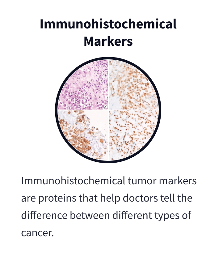
Immunohistochemistry is crucial for a mesothelioma diagnosis. IHC uses dyes that highlight proteins in tissue samples. These proteins act as markers of specific diseases. They can help doctors tell mesothelioma from lung cancer, for example, and distinguish between different mesothelioma subtypes.

What Is Immunohistochemistry?

Immunohistochemistry is a valuable tool for diagnosing mesothelioma. Its results are typically included as part of your pathology report. IHC involves staining tissue samples with a special dye and looking for signs of diseases like cancer.
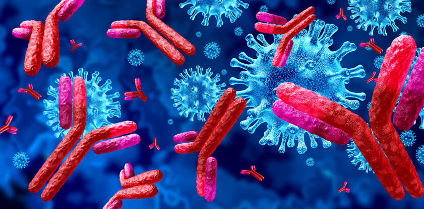
The dye used in IHC contains antibodies, which are proteins your immune system makes to fight illnesses. These antibodies can recognize and attach themselves to things that can harm you, such as viruses or bacteria. They can also attach to other proteins that can be signs of cancer or cancer markers.
Markers for mesothelioma include proteins such as calretinin, WT1 and cytokeratin 5/6. Identifying these key proteins helps pathologists confirm if someone has mesothelioma and which specific mesothelioma subtype they have.
Learn about your diagnosis, top doctors and how to pay for treatment in our free mesothelioma guide.
Get Your GuideWhy Are Immunohistochemical Markers Important for Mesothelioma Diagnosis?
IHC markers are a vital part of a mesothelioma diagnosis because they help ensure accuracy. An initial misdiagnosis is unfortunately common because mesothelioma can initially look like other cancers. Combining multiple markers enhances diagnostic precision and reduces misdiagnosis.
Dr. Snehal Smart, a mesothelioma specialist and Patient Advocate at The Mesothelioma Center, explains how IHC ensures patients get the correct treatment faster. “IHC markers help the pathologist distinguish between mesothelioma subtypes” she says, “This reduces the chances of misdiagnosis.”
Key Benefits of IHC for Mesothelioma
- Detect specific mesothelioma markers: Key markers include calretinin, WT1, cytokeratin 5/6, podoplanin (D2-40) and mesothelin.
- Differentiate subtypes: Identifies epithelioid, sarcomatoid and biphasic mesothelioma.
- Link to asbestos exposure: Confirms cancers related to asbestos.
IHC works alongside other tools like CT scans and genetic testing. Together, they help create a comprehensive diagnostic approach. A multidisciplinary team ensures accurate diagnosis and treatment planning.
As Snehal explains, “When patients call and ask to review their pathology report with me, I review their mesothelioma subtype and explain how this helps their doctor determine the best treatment plan for them. Subtypes respond differently to various mesothelioma treatments.”

“After reading the guide, I felt more confident about what was ahead.” – Carla F., mesothelioma survivor
Get Your Free GuideCommon Immunohistochemical Markers for Mesothelioma
Pathologists know to look for specific markers that are commonly associated with mesothelioma. Each marker confirms the disease and differentiates it from other cancers.
Key Markers for Mesothelioma:
- Calretinin: This protein is commonly present in mesothelioma cells, especially sarcomatoid cells.
- Cytokeratin 5/6: This protein is identified in more than 75% of mesothelioma cases.
- Mesothelin: Found in all mesothelioma cells, high levels of this protein indicate cancer and can help rule out other cancers.
- Podoplanin: Also known as D2-40, this protein differentiates mesothelioma from adenocarcinoma, a cancer that starts in the glands.
- WT1 protein: The WT-1 gene produces this protein that helps confirm pleural mesothelioma.
Additional markers may also become more commonly used to accurately diagnose mesothelioma. A 2021 study suggests the newly found MUC4 and GATA 3 markers help doctors distinguish between pleural sarcomatoid mesothelioma and pulmonary sarcomatoid carcinoma.
Comparing Mesothelioma Markers to Other Cancer Markers
Common mesothelioma markers or positive markers alone can’t rule out other cancers. Some markers are considered negative mesothelioma markers. They typically aren’t found in mesothelioma cells. If these negative mesothelioma markers are found in samples, the tumor may be a different type of cancer.
Common Negative Mesothelioma Markers
- B72.3
- CD15
- CDX2
- CEA
- MOC31
- Naspin A
- PAX2
- PAX8
- TTF-1
While some positive markers for mesothelioma are unique to the disease, others overlap with markers for other cancers. EMA, for example, is found in various cancers including mesothelioma. When markers aren’t exclusive to mesothelioma, it’s the whole picture of combined markers that can improve the accuracy of a diagnosis.
Cytokeratin is found in both mesothelioma and adenocarcinoma. However, if markers such as WT1 and calretinin are also found, this indicates mesothelioma. Combining markers improves diagnostic precision and reduces errors.
Common Questions About Immunohistochemical Markers for Mesothelioma
- What Are the Most Reliable IHC Markers for Mesothelioma?
-
Calretinin, WT1 and cytokeratin 5/6 are the most reliable markers for mesothelioma. They’re highly sensitive and specific.
- How Long Does IHC Testing Take?
-
IHC testing typically takes 1-3 days, depending on the lab’s processing time and the number of analyzed markers.
- Can IHC Markers Determine Mesothelioma Subtypes?
-
Yes, markers like calretinin and cytokeratin 5/6 help identify epithelioid or sarcomatoid mesothelioma subtypes.
- What Is the Role of IHC in Asbestos-Related Cancers?
-
IHC confirms asbestos-related cancers, such as mesothelioma. It identifies markers specific to asbestos-linked tumors.


