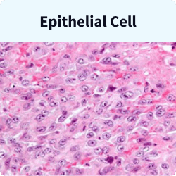
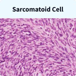
Mesothelioma Cells
Epithelioid, sarcomatoid and biphasic are the three main types of mesothelioma cells. Epithelioid is the most common type and offers the best prognosis. Meanwhile, sarcomatoid is rare and has a poor prognosis. Biphasic has a mix of both cell types, and prognosis depends on the ratio of each.
Key Facts About Mesothelioma Cells
- A biopsy is crucial for diagnosing mesothelioma. It alone can determine the specific mesothelioma cell type.
- Your tumor’s location and cell type will affect your treatment options.
- A study on peritoneal mesothelioma found a median survival of 55 months for patients with epithelioid cells. Patients with sarcomatoid or biphasic cells had a median survival of 7 to 13 months.
What Are the Different Types of Mesothelioma Cells?
There are three major mesothelioma cell types. These cell types differ in how they look under a microscope. They also differ in how they grow and form tumors in the body.
- Epithelioid: This is the most common mesothelioma cell type. It accounts for 50% to 70% of all cases. Epithelioid is the most responsive to treatment and has a better prognosis than other cell types.
- Sarcomatoid: This rare type of mesothelioma cell is spindle-shaped and has a poor prognosis.
- Biphasic: This cell type is a combination of epithelioid and sarcomatoid cells. Treatment options and prognosis depend on the ratio of each cell type.
Many rare cell subtypes exist within the three main mesothelioma cell categories. These subtypes have characteristics similar to those of the larger cell category they belong to.
Each cell type responds to treatment differently. The cell type also affects prognosis. After a mesothelioma diagnosis, your doctor will study your mesothelioma pathology report. This report helps them to understand the details of your cancer cell type.
This information is critical. Testing for the mesothelioma cell type is necessary to develop a helpful treatment plan.
What Is Mesothelioma Histology?
Mesothelioma histology is the study of cancerous mesothelial cells. Histopathology, a branch of pathology, studies diseased cells. A pathologist uses it to determine your cancer cell type. Yet only a few specialize in mesothelioma.
These specialists use microscopes to view cells up close. They prepare samples of tissue with chemical stains that make the cells’ features stand out. A 2021 research study suggests using electron microscopy for a more accurate mesothelioma diagnosis.
Histology also helps prevent misdiagnosis. It allows doctors to study cell types and distinguish between diseases. For instance, peritoneal mesothelioma and ovarian cancer can be hard to tell apart.

Cell Types of Malignant Mesothelioma
Pathologists look for three types of cells: epithelial, sarcomatoid and biphasic. Each cell type has different visible traits under a microscope.
For example, sarcomatoid cells have long nuclei. Epithelial cells have microvilli, microscopic protrusions of the cell. They also have clear structures called organelles within each cell. Doctors use these features to confirm the diagnosis.
The appearance of the different cell types is subtle. Only the most experienced pathologists can tell the difference. This nuance can make a diagnosis quite tricky. For example, it can be hard to tell mesothelioma cells from adenocarcinoma cells.
Epithelial Cells
Epithelial cells are uniform, sharply defined and shaped like squares or tubes. They have large nuclei and divide quickly. They often stick together, which hinders their spread in the body. Epithelial cells account 50% to 70% of mesothelioma cases.
Epithelial mesothelioma usually responds best to treatment. This can lead to a better prognosis
Sarcomatoid Cells
Spindle-shaped sarcomatoid cells lack a defined structure and are irregularly shaped. They spread faster than epithelial cells because they don’t stick together. These cells only account for 10% to 20% of cases.
Sarcomatoid cell mesothelioma is more aggressive and often diagnosed later than the epithelial type. This leads to fewer treatment options. Its tumors are less defined, making surgery harder. As a result, some mesothelioma specialists do not consider sarcomatoid tumors for surgery.
Biphasic Cells
Mesothelioma is biphasic when it contains epithelial and sarcomatoid cells. This mixed cell type accounts for 20% to 30% of mesothelioma cases. Each cell type must account for at least 10% of the tumor mass to receive a biphasic diagnosis.
Treatment options are better and life expectancy is longer if there are more epithelial cells and fewer sarcomatoid cells. Treatment options can include surgery, immunotherapy, chemotherapy and radiation.
Doctors use cell type and mesothelioma staging to estimate prognosis and develop a treatment plan. When treatment targets the exact cell type, patients may live longer.
Rare Variances in Histological Mesothelioma Types
Some rare mesothelioma cells require a more detailed classification within the three main cell types.
Adenomatoid
In this variant of epithelial mesothelioma, the cells line small, gland-like structures. This type is also called glandular or microglandular mesothelioma.
Benign
Benign mesothelioma is neither cancerous nor the result of asbestos exposure.
Cystic
The cystic mesothelioma type has smooth, thin-walled cysts held together by fragile fibrovascular tissue. It is a subtype of epithelial mesothelioma.
Deciduoid
Deciduoid reflects this unusual epithelial cell subtype’s histological resemblance to cell changes occurring in early pregnancy. It most commonly affects young women.
Desmoplastic
Desmoplastic mesothelioma is a form of sarcomatoid mesothelioma, and more than 50% of the tumor comprises dense, fibrous tissue.
Heterologous
Tumors of heterologous cell types contain bodily tissues different from those in which they form. Only a handful of cases exist in the medical literature.
Lymphohistiocytoid
This subtype of sarcomatoid mesothelioma, known as lymphohistiocytoid mesothelioma, often leads to misdiagnosis. Its makeup resembles a dense bundle of inflammatory and immune cells.
Papillary
This variant of epithelial mesothelioma, known as papillary mesothelioma, resembles healthy cells that grow and multiply slowly. It does not typically spread to other parts of the body.
Small Cell
Small cell mesothelioma occurs when a large proportion of a mesothelioma tumor comprises small cells that grow in a pattern similar to small-cell carcinoma.
Histology Process to Determine Mesothelioma Cell Type
The histology process starts with the surgeon who takes the biopsy. A technician transfers the tissue from the biopsy onto a slide for a microscope. The pathologist then works to identify cancer cells.

Surgeons remove tumor tissue during a biopsy. In the lab, a technician fixes, embeds, sections and mounts the cells on a slide. Then, chemical stains reveal the cells’ appearance for a pathologist to identify.
Once the technician mounts and stains the biopsy cells on a slide, the sample is ready to view under a microscope. The pathologist notes the cells’ size, shape and structure to identify the tumor’s cell type.
A frozen section fixation is used to diagnose cancer during surgery. Small tumor pieces are frozen while the patient is still in surgery. Then, a pathologist stains and examines a slice of this tissue. This quick check reveals if the tumor is malignant.
Additional Lab Processes to Support Histology Cell Studies
Pathologists use other techniques to learn more about cells. These techniques include in situ hybridization and immunohistochemistry.
- In situ hybridization: This technique uses fluorescent or radioactive probes to bind DNA and RNA. Scientists can use this method to analyze a cell’s genes and detect genetic abnormalities.
- Immunohistochemistry: This technique relies on the principle that antibodies bind to specific antigens. Antibodies also bind to cancer cell proteins called oncoproteins. Depending on the suspected cancer type, antibodies are applied to tissues on a microscope slide. The interaction of antibodies and oncoproteins creates visual patterns, which help pathologists diagnose mesothelioma.
Common immunohistochemical markers for mesothelioma include calretinin and cytokeratin 5 and 6. Pathologists also look for the WT-1 protein and podoplanin (D2-40). Immunohistochemistry is often combined with other tests to offer the most accurate diagnosis of mesothelioma

Why Is Mesothelioma Pathology Important?
Pathology tests are vital to confirming a mesothelioma diagnosis. This testing helps doctors determine the best treatment plans.
The pathology report is the most crucial component in a mesothelioma diagnosis. It provides the definitive cancer diagnosis, describing the specific types of cells found in a patient’s tumors.
Doctors recommend more aggressive treatment for patients with epithelial cells because they typically respond better. Doctors also consider other factors when developing treatment plans. These factors include the stage of the cancer’s progression and the patient’s age, gender and overall health.
Recommended Reading


